Rudinas – Zarate, Catheryn Rose, MD, Toring, John Concordio, MD, Lagula – Bilocura, Imelda Ramirez, MD
Long-standing, uncontrolled hyperthyroidism is commonly associated with cardiac complications, including dilated cardiomyopathy, congestive heart failure and cardiac arrhythmias. The association of hyperthyroidism with elevated troponins and/or myocarditis however, is scarcely reported, more notably in the young. Here we present 2 patients, both recently diagnosed with Grave’s disease, presenting at the emergency room with acute onset of cardiac symptoms:
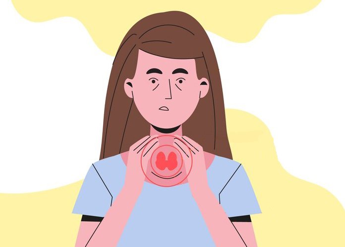
Case 1. A 26-year-old female presented with palpitations, chest pain radiating to the left arm and difficulty breathing. There was no recent history of cough, colds and fever. Her past medical history and family history were unremarkable. She had no vices and was unemployed at that time. Her vital signs were normal except for tachycardia at 139bpm, with regular rhythm. Pertinent physical examination findings included mild agitation, bilateral exophthalmos, resting tremors and an easily palpable and visible thyroid gland with no distinct nodules.
Electrocardiogram showed sinus tachycardia, anterolateral and inferior wall ischemia with incomplete right bundle branch block. Troponin I was noted to be elevated at 576.5 ng/L (reference range <19 ng/L. Echocardiography showed normal left ventricular geometry with normal biventricular resting systolic and diastolic functions. TSH was suppressed at 0.006 uIU/ mL (reference range 0.3 – 5.0 uIU/mL), FT4 was elevated at 88.08 pmol/L (reference range 11.0-22.5 pmol/L) and FT3 was high at 39.15 pmol/L (reference range 3.1-6.5 pmol/L). Liver enzymes, SGPT and SGOT were noted to be mildly elevated at 56 U/L (reference range 5 – 50 U/L) and 42 U/L (reference range 10 – 40 U/L), respectively. Ultrasound of the neck showed an enlarged thyroid gland with diffusely coarsened parenchyma, with doppler evidence of increased vascularity. TRab and Anti-TPO antibodies were both positive.
The patient was managed as a case of Grave’s disease in Impending Thyroid Storm (Burch-Wartofsky score of 30) and Probable Acute Myocarditis. Propylthiouracil 200 mg every 6 hours and propanolol 40 mg every 8 hours were started along with aspirin, colchicine and high-dose statin. On the 2nd day of admission, troponin I was noted to further increase to 1019.5 ng/L (reference range <19 ng/L). Cardiac MRI and calcium scoring were requested by cardio service but were not performed due to financial limitations. On the subsequent hospital days, her chest pain improved and eventually resolved. The palpitations, tremors and agitation resolved as well. On the 5th hospital day, repeat Troponin I was significantly lower at 107.1 ng/L (reference range <19 ng/L), although it was still above the normal value. She was eventually discharged with methimazole, propanolol, aspirin, colchicine and atorvastatin.
On follow-up after 3 months, the patient was stable with no recurrence of the previously mentioned symptoms. Repeat TSH, FT4, FT3, SGPT, Troponin I and electrocardiography were all within normal.

Case 2. A 24-year-old male presented with an 8-hour history of bilateral lower extremity weakness following a wine drinking session with friends. Symptoms of chest pain and dyspnea noted 2 hours prior to admission prompted emergent consult. He had no previous comorbidities and was not on any maintenance medications.
At the emergency department, the patient complained of mild chest discomfort, associated with dyspnea and palpitations. A markedly elevated heart rate approaching 160 bpm was noted, and a subsequent electrocardiogram revealed supraventricular tachycardia. The rest of her vital signs were within normal limits. Pharmacologic cardioversion was done, with rhythm reverting to sinus. Notable laboratories at this point included a markedly decreased potassium level of 1.1 mmol/L, suppressed TSH of <0.005 uIU/mL (reference range 0.3 – 5.0 uIU/mL), elevated thyroid hormones of FT4 at 54.19 pmol/L (reference range 11.0-22.5 pmol/L) and FT3 at 21.86 pmol/L (reference range 3.1-6.5 pmol/L), and a markedly elevated Troponin I level of 320.4 ng/L (reference range <19 ng/L), with a repeat value of 800 ng/L.
The patient was managed as a case of Thyrotoxic Heart Disease with Cardiac Dysrhythmia: SVT, converted to Sinus Rhythm, Primary Hyperthyroidism secondary to Graves’ Disease, and Thyrotoxic Periodic Paralysis. Methimazole at a total dose of 30 mg daily was started, along with Propranolol 40 mg given every 8 hours. Cautious K replacement was done. Aspirin and atorvastatin were likewise started. Neck ultrasonography revealed globularly enlarged thyroid gland with diffuse parenchymal changes with micronodulations and doppler evidence of slightly increased vascularity, consistent with a hyperthyroid state. Echocardiography revealed normal left ventricular geometry with multisegmental wall motion abnormalities and a mildly reduced left ventricular systolic function (45%), with doppler evidence of grade 3 diastolic dysfunction.
Sustained clinical improvement was seen during the remainder of the clinical course, with normalization of serum potassium and resolution of bilateral extremity weakness, chest pain, and dyspnea. The patient was discharged with methimazole, propranolol, aspirin, and atorvastatin.
On follow-up at 2 weeks, the patient reported no recurrence of the previous symptoms. Repeat laboratory tests revealed a decreasing level of thyroid hormones with FT4 at 44.45 pmol/L and FT3 at 15.58 pmol/L. Troponin-I level at that time was normal at 6 ng/L.
In summary, we have presented two young patients, both diagnosed with Grave’s disease during their initial consultation at the emergency room. Both had a low pretest probability of having coronary artery disease, although both manifested with an acute onset of angina and markedly elevated Troponin I levels. A similar case has been cited by Lancaster (2) of a 29-year-old patient with hyperthyroidism, elevated thyroxine, suppressed TSH, and acute onset of cardiac symptoms with elevated cardiac enzymes. Cardiac magnetic resonance imaging (CMR) was performed, demonstrating diffuse left ventricular edema and subepicardial late gadolinium enhancement in the mid‐to‐distal anterior and anterolateral walls – consistent with the clinical picture of acute myocarditis. The same patient was started on methimazole and on follow-up, resolution of cardiac symptoms was noted, along with control of hyperthyroidism. In a case series by Fatourechi (1), eleven (11) patients with Graves’ disease and unexplained systolic dysfunction were subjected to endomyocardial biopsy. Results showed the presence of lymphocytic infiltrates in two (2) of the biopsies consistent with autoimmunity. Literature is limited regarding these cases, and up to now, there is still no consensus on the exact mechanism underlying the autoimmune myocarditis that can sometimes be seen in patients with Graves’ Disease. Moreover, screening all hyperthyroid patients with cardiac magnetic resonance imaging (CMR) and performing endomyocardial biopsy to investigate for myocarditis is cost prohibitive and unnecessarily invasive with regards to the latter.
As endocrinologists at the forefront in the care of patients with hyperthyroidism, where does this take us? How often do we get cardiac biomarkers for our young hyperthyroid patients with chest discomfort? Would we want to check cardiac biomarkers for our hyperthyroid patients without cardiac symptoms as well? In terms of management, what then is the role of antiplatelets and high-dose statins? If autoimmunity in fact plays a central role in the pathogenesis, what is the role of steroids? Lastly, will colchicine, with its anti-inflammatory and cardioprotective effects, benefit these patients?
The world of endocrinology is indeed a wonderful and mysterious one, and we leave these things and more for you to ponder upon.
References
- Fatourechi V, Edwards WD. Graves’ disease and low‐output cardiac dysfunction: implications for autoimmune disease in endomyocardial biopsy tissue from eleven patients. Thyroid. 2000;10(7):601‐605.
- Lancaster ST, Koons KL, Lee YJ, Mazimba S, Kwon Y. Acute autoimmune myocarditis as a manifestation of Graves’ disease: A case report and review of the literature. Clin Case Rep. 2019;7:1489–1493. 10.1002/ccr3.2273
- Mavrogeni S, Markussis V, Bratis K, et al. Hyperthyroidism induced autoimmune myocarditis. Evaluation by cardiovascular magnetic resonance and endomyocardial biopsy. Int J Cardiol. 2012;158:166‐168.